

_(14735220116).jpg)
The term “health physics” was first coined in the University of Chicago’s Metallurgical Laboratory during the early years of the Manhattan Project. As scientists converged on Chicago during World War II to study the potential of nuclear energy, some physicists turned their attention to learning how to protect people from radiation. Meanwhile, physicians capitalized on the growing production of radioactive products to investigate novel treatments for disease. Health physics and the related field of nuclear medicine, however, have a much longer history.
Beginnings
Radiation’s potential as a medical tool was recognized very quickly after its discovery. The x-ray, discovered by Wilhelm Conrad Roentgen in 1895, was adopted by physicians immediately to photograph the bones beneath the skin’s surface. Shortly after the discovery of radioactivity by Henri Becquerel in 1896, it was suggested independently by both Pierre Curie and Alexander Graham Bell that a radioactive source inserted into a tumor could cause the tumor to shrink. Research began in the early twentieth century into methods for applying localized radiation to tumors.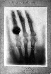
The potential harm of radiation also quickly became apparent, and procedures were put into place to try and limit its damage. In 1898, the Roentgen Society created a committee on x-ray injuries. A Boston dentist named William Herbert Rollins performed experiments on guinea pigs, which showed that x-rays could kill unborn babies as well as adult guinea pigs. Rollins used leaded tube housings to limit radiation, and advised against x-rays in exams of pregnant women. At a meeting of the American Roentgen Ray Society, an x-ray tube manufacturer described his practice of carrying a photographic plate with him, which he would develop each night to see if he had been exposed to radiation. While not adopted, this practice predated the modern film badge by a number of years.
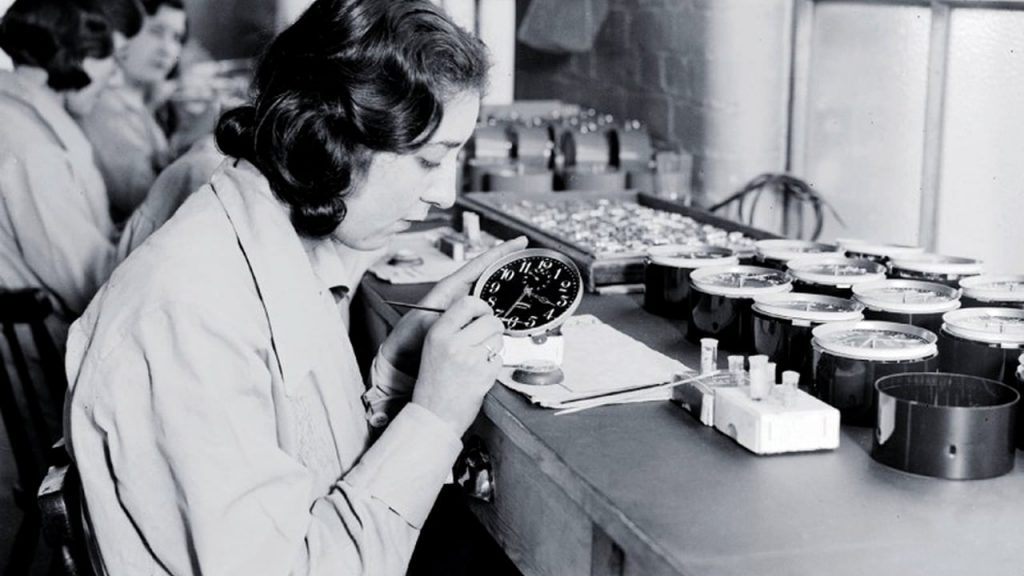
The extent of the damage radiation could cause, however, was still not known. In fact, many popular products included radioactive materials. Radium, for instance, was used in medicines, cosmetics, and in paint for watch dials, exposing those who handled it to harmful radiation. Once more information was uncovered about the effects of radiation, these products went out of style and the implementation of security measures gained momentum.
The 1920s saw a large number of advances in radiation safety. Radiation protection recommendations were adopted by the British Roentgen Society in 1915 and the American Roentgen Ray Society in 1922. The first permissible exposure limit, which marked the highest dose considered safe at the time, was set by physicist Arthur Mutscheller at a meeting of the American Roentgen Ray Society in 1924. He set the limit to be 1/100 of the amount of radiation known to cause a reddening of the skin, called erythema, in a month. Other innovations included Hermann Muller’s discovery that high-energy radiation produces genetic mutations; the establishment of the roentgen unit of radiation; and the adoption of the film badge, a small device containing photographic film that measured radiation exposure levels. During the 1930s, the permissible exposure limit was lowered a number of times.
Nuclear medicine also has its foundations early in the twentieth century. In 1911, Hungarian Physicist George de Hevesy realized that lead could be tagged with radioactive radium. In the 1920s, de Hevesy experimented with labeled atoms to probe animal and plant physiology. Shortly after deuterium was discovered in the early 1930s, de Hevesy also developed the first clinical use of isotopes in his studies of water content in the human body. He received the Nobel Prize in Chemistry in 1943 “for his work on the use of isotopes as tracers in the study of chemical processes.”
Berkeley
Many claim that the “genesis” of the field of nuclear medicine occurred at Berkeley, when physician John Lawrence came to the Radiation Laboratory at the request of his brother, physicist Ernest Lawrence. While research into the subject did not start at Berkeley, the program undertaken by John Lawrence and his team was unprecedented in its scope and results.
John Lawrence’s group, which would become the Donner Laboratory, made use of radioactive materials created in Ernest Lawrence’s cyclotron. The lab’s main focus was in the use of tracers, or complexes made up of radioactive material and biologically relevant molecules, for studying metabolism and for treating patients. This included using phosphorous-32 to treat leukemia, marking the first time a radioactive isotope was used to treat a disease. The lab also discovered several new radioactive isotopes, called radioisotopes, that would later become common in nuclear medicine, including technetium-99m, carbon-14, and fluorine-18.
The Berkeley Radiation Lab thus became the center of studies into the medical benefits of radioisotopes, even before the Manhattan Project started. In addition to John Lawrence’s work, Joseph Hamilton turned his attention toward radioactive iodine, which localized to the thyroid glands. Physicians Robert Stone and Paul Aebersold investigated the 60-inch cyclotron as a neutron therapy source, bombarding patients with neutrons to initially positive results. A true testament to the Lawrences’ belief in their new treatments, the first patient of the neutron therapy was John and Ernest Lawrence’s mother, who was suffering from cancer in her pelvis. After her treatment, she lived another eighteen years. While neutrons were effective at shrinking tumors, they were also hard to control. Some of the patients experienced painful side effects.
Leading Up to the Manhattan Project
Research into health physics intensified during the Manhattan Project. As secret cities rose out of the mountains and valleys of Los Alamos, Hanford, and Oak Ridge, burning questions still remained as to how to protect the residents from the radioactive products that were used at each site. While research into health physics sped up during the war, nuclear medicine came back into vogue after the war ended, carried by the wave of radioactive isotopes being produced at former Manhattan Project sites around the country.